Diagnostic Imaging

Diagnostic Imaging
Our imaging and radiology services teams work with doctors, nurses, and other clinicians to deliver comprehensive care. Tests include:
-
Angiography: Used to examine the inside of arteries and veins to detect blockages or narrowing of the vessels. Evaluates blood flow, or detects brain aneurysms or blood vessel abnormalities. It is used to visualize renal, carotid and vertebral arteries, or examine the aorta for aneurysm.
-
Biopsy: To determine whether a growth is cancerous, a tissue sample is extracted for further testing. Often used in diagnosing cancer when a mass is found during a screening exam. Can be performed by needle, stereotactic or surgical.
-
Bone Densitometry (DEXA (Dual Energy X-Ray Absorptiometry): Measures the strength of bones to determine if there is risk for developing osteoporosis or osteopenia (decreased bone mass). The test is used to develop an appropriate treatment plan to slow the progression of disease and prevent fractures.
-
Although both men and women can develop osteoporosis, women are at greater risk for developing the disease, particularly after menopause.
-
-
Breast Imaging:
-
Digital mammography is an X-ray exam of the breasts used to screen for or diagnose breast cancer. With digital technology, radiologists can zoom in on particular areas or change brightness or contrast for even greater visibility, and results can be read immediately.
-
Breast MRI is often used for women who are at greater risk of developing breast cancer or who have dense breast tissue or implants - cases in which mammography is less effective at detecting abnormalities. This technique is used to assess the extent of breast cancer, determine the effectiveness of chemotherapy or radiation therapy, further evaluate abnormalities and provide additional detail for treatment planning.
-
Breast ultrasound is often used to further evaluate an abnormality found during a mammogram. Ultrasound allows doctors to see the area closest to the chest wall, which can be difficult to see using mammography. This technology also helps doctors determine whether a breast lump is filled with fluid (a cyst) or is a solid mass.
-
Digital stereotactic biopsies, and women can choose to be seated or lying down during the procedure for maximum comfort.
-
Cardiac Catheterization: Used to guide small wires or catheters (thin, flexible tube) with specialized instruments to treat affected areas of the body. These procedures only require a tiny incision where the catheter is inserted into an artery, so it results in less blood loss, less pain and a quicker recovery for patients.
-
Advanced 64-slice CT Technology: Can capture images of a beating heart in five heartbeats, an organ in one second, and perform a whole body scan in 10 seconds that results in less radiation exposure for patients, and can be used to examine a wider range of conditions.
-
CT: (computed tomography or CAT) scan combines X-ray and computer technology to show highly detailed, 3-D images of any part of the body, including bones, muscles, fat, organs and blood vessels. Scans can also be performed using a contrast solution (either swallowed or injected) to make tissues and vessels more visible.
-
Fluoroscopy: Uses X-rays to provide real-time images of the area being examined, often to examine various body systems, including skeletal, digestive, urinary or reproductive, as well as organs such as the heart, lungs and kidneys. Fluoroscopy is commonly used to examine the intestines and large bowel, and is most often performed using a contrast solution to make tissues and other structures more visible.
-
Interventional Radiology: We offer a range of minimally invasive interventional radiology techniques to offer safe and effective options to treat a variety of conditions, including angiography, embolization, radiofrequency ablation, vertebroplasty and cardiac catheterization.
-
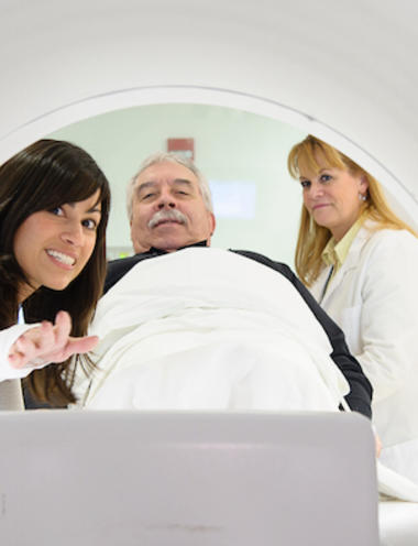
MRI: Magnetic resonance imaging combines a powerful magnet, radio waves and computer technology to provide detailed images of tissues, muscles, nerves and bones. Because MRI uses magnetic force and radio waves to create images, there is no radiation exposure during the procedure.
-
Nuclear Medicine: Uses very small amounts of radioactive materials (given either orally or intravenously) to examine an organ's structure and metabolic function, and is used to scan organs for abnormalities, evaluate the spread of cancer, locate infection and identify blood clots in the lungs.
-
PET/CT: Provide specific information about organ and cell functioning by distinguishing among healthy, diseased and dead tissue. Our PET/CT scanning technology combines the physiological information from a PET scan and the anatomical information from a CT scan to provide a comprehensive image of the body in a single scan.
-
Ultrasound: Uses reflected sound waves to create real-time images of soft tissues, including muscles, blood vessels and organs, causing no radiation exposure during this procedure. Most commonly used to examine the fetus during pregnancy, it is also an effective tool for monitoring blood flow using Doppler ultrasound technology. Can also be used to discover abnormalities in organs, and detect narrowed arteries, clotted veins, or growths such as tumors and cysts.
-
X-ray: Uses invisible electromagnetic energy beams to produce images of internal tissues, bones and organs on film or digital media. Performed for many reasons, including diagnosing tumors or bone injuries and for procedures, such as arteriograms, computed tomography (CT) scans and fluoroscopy.
We accept walk-in patients for X-ray and lab orders with a prescription.
Monday - Friday, 7 a.m. - 5 p.m.
13695 US Highway 1
Sebastian, FL 32958
772-589-3186
